How can you assess the risk of a mole in just two minutes—or diagnose a complex disease from a single drop of blood? How are engineers and digital technologies supporting doctors today—and vice versa? We spoke with Irina Matveyeva, a biotech engineer and lecturer at Samara University, to find out.
— Irina, what exactly does a biotech engineer do?
I specialize in medical technology, applying various engineering methods to medicine. I work at the Department of Laser and Biotechnical Systems, where we train engineers, electronics specialists, and IT professionals who deeply understand medicine and biology—and who know how to apply engineering solutions in these fields. Our focus is on developing medical devices that incorporate laser and optical technologies.
— Can you give an example?
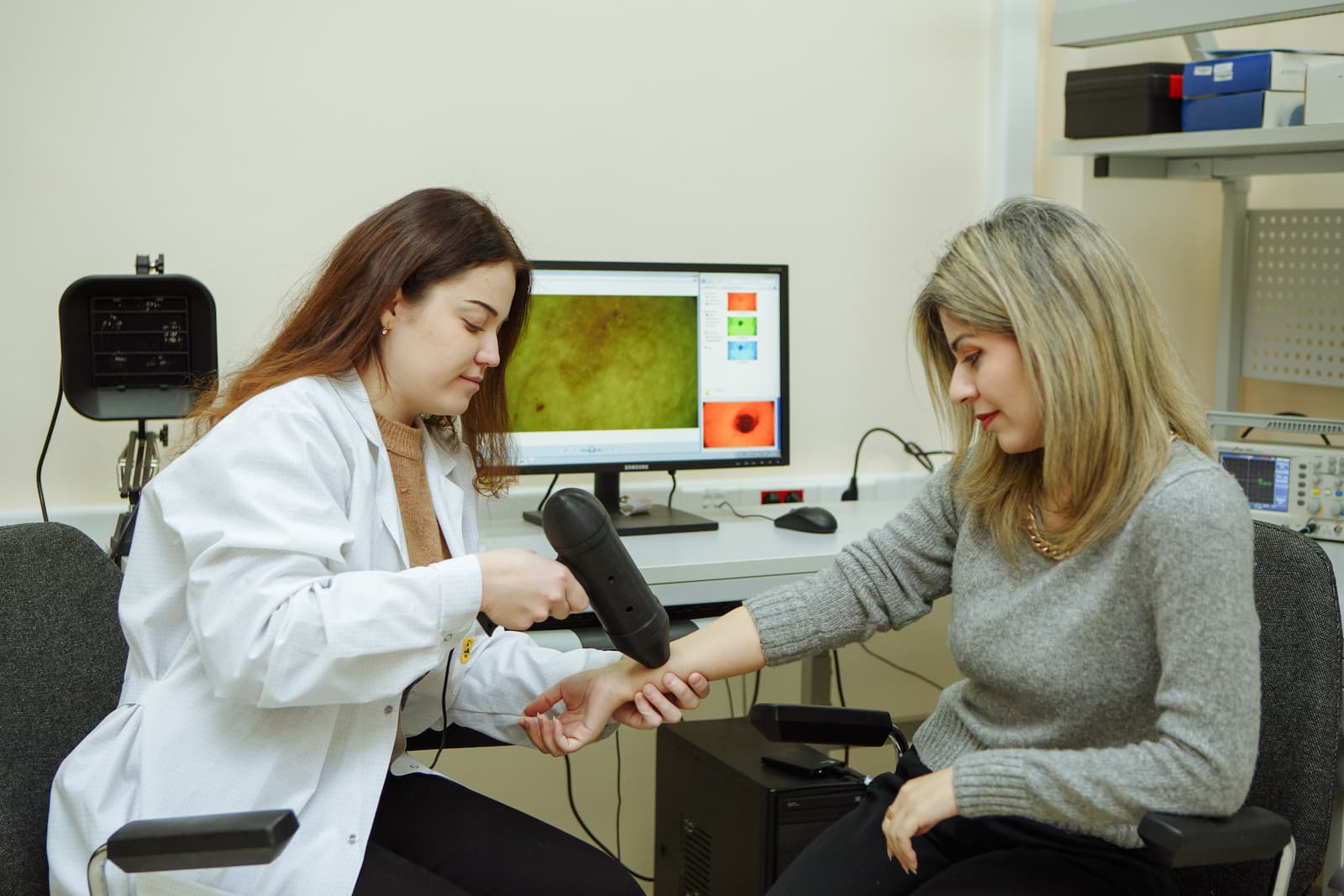
One of our key developments is a multimodal diagnostic system comprising two devices designed to evaluate moles and detect malignant skin lesions.
The first component is a Raman spectrometer—a device that captures light scattered in a very specific way. In Raman spectroscopy, light exchanges energy with molecules in the sample. By analyzing this interaction, we can determine the biochemical characteristics of a skin lesion. How do we collect this data? We simply shine a laser on the mole for two minutes—completely painlessly and non-invasively.
The second part is essentially a high-resolution imaging system: a specialized camera that captures detailed images of the mole, allowing us to assess its external morphological features.
Both sets of data—spectral and visual—are then analyzed by a neural network trained on a vast “library” of previously identified patterns. Within seconds, the system delivers a verdict: benign or potentially dangerous.
This approach is entirely non-invasive. Traditionally, if a mole raises suspicion, it’s surgically removed and examined under a microscope. Our method, however, can predict malignancy with up to 95% accuracy—nearly as precise as histological analysis, yet far more efficient than a standard visual examination by a physician.
— How do you train the artificial intelligence?
We use anonymized clinical data from patients whose malignant lesions were surgically removed, in close collaboration with the Samara Regional Oncology Dispensary.
— What stage is your development at now?
We’ve filed a patent application for the device. It’s already being used at the Samara Oncology Center as a supplementary diagnostic tool. To move forward with full-scale clinical trials, we need to gather more patient data and refine our analytical algorithms—a process we expect to take about another year.
— I’ve heard you’re also working on cancer detection from a drop of blood.
Yes, we use the same Raman spectrometer for that. We direct a laser beam onto literally a single drop of blood and obtain a molecular spectrum that can reveal not only oncological conditions—such as multiple myeloma—but also other serious diseases like heart failure, kidney dysfunction, and systemic lupus erythematosus.
Again, a neural network performs complex calculations in real time and delivers a diagnosis with 75–95% accuracy. And unlike conventional lab tests, this method doesn’t require multiple vials of blood, expensive reagents, specialized lab equipment, or highly trained technicians. All you need is a compact laser device powered by electricity and a software program. Crucially, this test could be performed in any local clinic—not just in specialized centers.
We see this as a future complementary diagnostic tool. Right now, we’re intensively training the AI model with help from clinicians at Samara State Medical University and the Regional Oncology Dispensary, who provide real patient data. The more data we collect—from thousands of patients—the better the AI learns complex disease patterns, and the higher the diagnostic accuracy becomes.
— So, in theory, could almost any disease be diagnosed this way—provided there’s enough training data?
Essentially, yes—if the disease produces detectable molecular changes in the sample. For instance, we recently began collaborating with ENT specialists from a Moscow medical center. They use our Raman spectrometer to scan tissue samples affected by various ear, nose, and throat conditions, then send the spectral data to us. Our students and postgraduates are now learning to identify ENT pathologies based on these spectra.
The methodology is proven. Now, we’re expanding our scope—studying as many different diseases as possible—to bring these technologies into broader areas of medicine.
Photos by Olesya Orina, Yulia Litvinova
Source: rg.ru