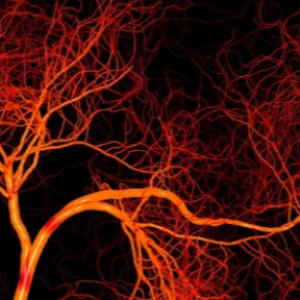
The effective and reliable digital image processing algorithm aimed at improving the visibility of veins has been created by researchers from Samara University. On the scientists’ plan, the development will help doctors more accurately insert a needle into a vein during medical procedures, especially in the case when the vein is not distinctly visible. The results are published in Journal of Biomedical Photonics & Engineering.
Modern methods of medical laboratory diagnostics often involve inserting a needle into a patient’s vein. However, sometimes the blood vessels are not distinctly visible, and performing this medical procedure causes difficulties for health workers, for example, it may be difficult to draw blood.
At present, for solving this problem, scientists actively developing the use of various optical methods, including near-infrared imaging of veins (similar to a night vision surveillance camera). However, these methods face a number of limitations, such as low depth and low contrast of the image. Besides, the digital image processing algorithms used in these methods are imperfect, having low stability and performance.
Scientists from Samara University have developed the efficient and reliable digital image processing algorithm for improving the visualization of veins. As Nikita Remizov, a graduate student at Samara University’s Department of Laser and Biotechnical Systems, informed us, the algorithm is built on efficient operations based on the discrete Fourier transform (an operation used in signal digitization algorithms, modifications of which are used, for example, in compressing audio to MP3 or images to JPEG).
“Image processing in Fourier space makes it possible to efficiently and reliably enhance areas in the image with sharp changes in the intensity of neighbouring pixels. The areas located in the high-frequency field of the two-dimensional spectrum correspond to sharp transitions in the image. Due to so-called fast Fourier transform, we can use relatively low-cost computing modules and process images in real time”, he said.
According to the researchers, the development surpasses current methods in terms of the efficiency of separating pixels corresponding to veins from pixels associated with surrounding body tissues.
“The new algorithm has become one of the stages in developing a domestic device for vein imaging. The device must be affordable in production, including that in conditions of the sanctions, and be effective and convenient for doctors when interacting with patients having a high body mass index and other factors that make venipuncture difficult”, said Remizov.
Today, scientists are optimizing the optical configuration of the future device. Combined with the algorithmic image processing, it will provide visualization of veins, which is not available when other methods are used, for example, in cases of severe skin pigmentation.
Source: ria.ru
Image: depositphotos.com
 RU
RU  EN
EN  CN
CN  ES
ES 