 RU
RU  EN
EN  CN
CN  ES
ES 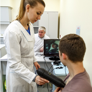
A team of scholars of Samara National Research University and Samara State Medical University as well as clinic specialists of Samara Regional Oncology Centre (SROC) tested a new way of skin cancer early diagnostics with the help of original complex of three devices.
The efficiency of oncology treatment is directly connected with the timeliness of their detection. For instance, for melanoma – the most aggressive form of skin tumour detected at the first stage – five-year survival rate after the treatment makes 98% and does not exceed 15% with diagnostics at the fourth stage.
Methods created by the team of Samara scholars allowed enhancing the efficiency of active and early skin neoplasm diagnostics up to 97% when the exactness of usual standard clinical research does not exceed 50-60%.
The research results are given in the article "Combined Raman and autofluorescence ex vivo diagnostics of skin cancer in NIR and visible regions" which was published in one of the most authoritative journals devoted to the use of modern optical technology for research in Medicine and Biology - Journal of Biomedical Optics1.
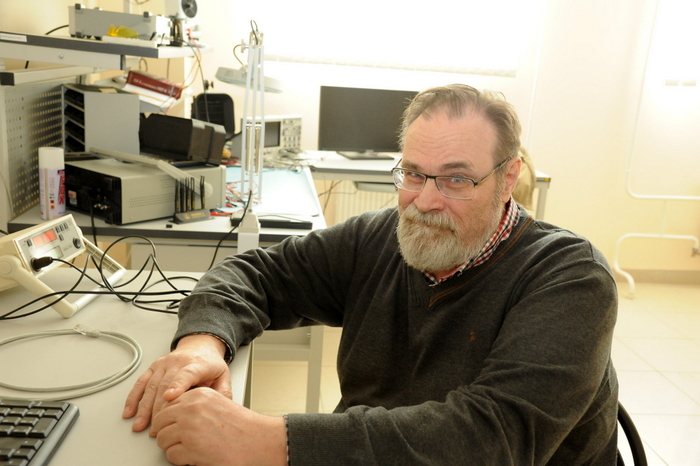
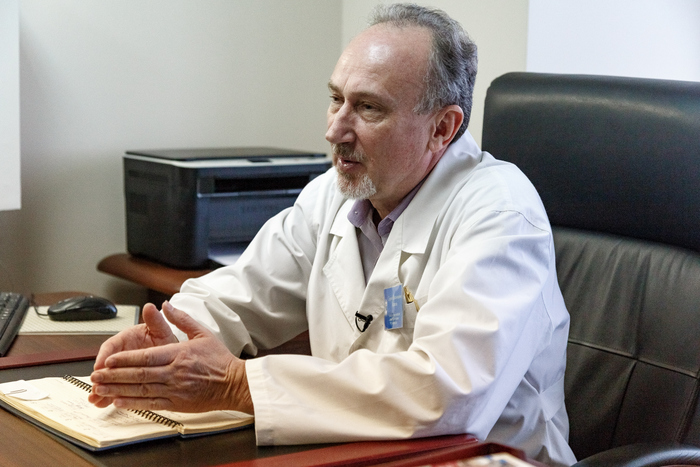
In the basis of methodology developed by Samara scholars at the Department of Laser and Biotechnical Systems of Samara University (headed by professor Valeriy Zakharov) is a system of spectral measurement of problem skin areas with the further medical interpretation of the received data offered by the Department of Oncology of SSMU (headed by professor Sergey Kozlov).
As professor Zakharov explained the developed system of spectral measurement allows registering parameters of a patient’s skin in many spectral ranges simultaneously and this gives opportunity to spot pathological changes on cell level as ill cells in their range are different from the healthy ones.
This means is the most suitable for screening programmes: not only it allows diagnosing pathology rather precisely and quickly, but it also does not require any use of additional consumable materials and chemical agents and permits to do without the use of invasive method of diagnosis confirmation. “For example, in case of suspicion in skin melanoma incorrect biopsy of tumour increases the risk of metastasis”, - Alexander Moryatov, the curator of the project, associate professor of the department of Oncology of SSMU added. – In any case after examination of a patient and surgical treatment we finally confirm the diagnosis morphologically”.
The scholars use three devices for diagnostics.
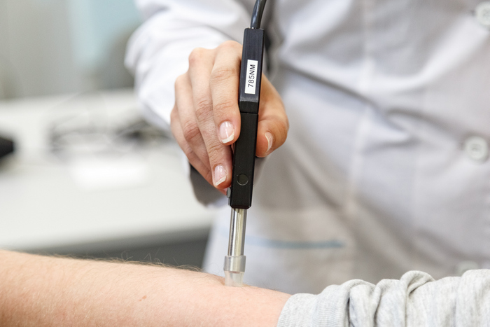
The first one is an experimental setting for measurement of Raman scattering, it studies spectral characteristics of neoplasms, first of all, skin melanoma. According to Sergey Kozlov this device is suitable for precise diagnostics of a malignant tumour nature allowing to diagnose and differentiate between melanoma and other forms of skin tumour quickly and safely. “This setting can be set in an ordinary regional hospital”, - the professor specified.
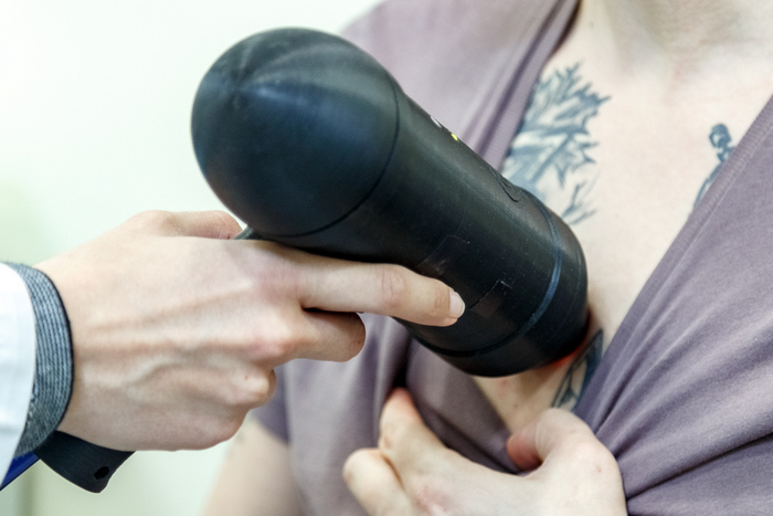
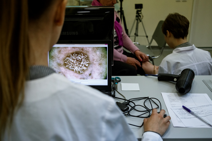
The second device – dermatoscope – gives a visual picture of tumour with the maximum approach to its surface and allows seeing and fixing characteristic signs of this disease in real time. According to Sergey Kozlov this device can be compared favourably with the similar ones in respect of a user-friendly interface, the ability to conduct analysis in the mode of polarized light, as well as with the help of special highlight to study peculiarities of melanin, hemoglobin, structure of capillary network in the studied skin area.
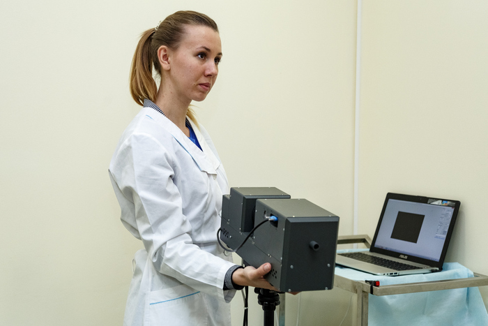
The third - hyperspectral camera – allows in a short period of time to make several tens of shots in different ranges (for different skin neoplasms are characteristic different optical characteristics) with high definition.
All three devices represent a joint development of Samara University scholars, SSMU and SROC and are directed at comparative research of optical characteristics of healthy and pathologically changed tissues in which analysis of different types of neoplasms is built on the basis of joint interpretation of their ranges got with the application of completely safe technologies and laser light sources of different wavelengths.
It is said about two types of spectral analysis the character of which depends on biochemical composition of tissues under investigation - Raman spectrum (RS)2 and autofluorescence spectrum (AS)3. “Thus “double” spectroscopy allows, as our experiments show, getting information about tissue structure on the basis of which different types of skin tumour can be identified rather precisely”, - Ivan Bratchenko, associate professor of Department of Laser and Biotechnical Systems of Samara University, asserts.
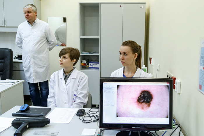
All three devices work at the department of Oncology of SSMU, clinical site of Samara Regional Clinical Ontology Dispensary and have already been used for several months when oncologists examine patients. With the help of this equipment more than 400 people have already been examined. “The received results give hope that we approach precise diagnostics comparable with morphological research”, - Alexander Moryatov mentioned.
According to Sergey Kozlov diagnostic devices showed high resolution and with time can emerge in the market of medical equipment: “We have few rivalries in Russia and abroad and here we have received better results”.
Samara scholars connect perspectives of their research in diagnostics of skin neoplasm with the increase of its resolution due to the complication of mathematical analysis of spectrum data. Their further plans are to integrate a complex of three devices with fiber-optics to use them for diagnosis of inner located tumours (gastrointestinal tract, lungs, etc.) preserving with this safe for a human being research principle – spectral analysis of tumours.
___________________________________
1Journal of Biomedical Optics is indexed in Scopus database since 1996 (to present time). According to the data of SCImago Journal Rank, the journal has the highest quartile Q1 in all scientific spheres, defined in the base: Atomic and Molecular Physic, and Optics; Biomaterials; Biomedical Engineering; Electronic, Optical and Magnetic Materials.
The editorial board of the journal includes scholars from the universities of the USA, Ireland, Sweden, Russia, Denmark, Great Britain, Austria, Italy, Switzerland, Australia, Germany, Canada, Norway, Taiwan and Hong Kong.
2Raman scattering (Raman effect) is the inelastic scattering of a photon by molecules which are excited to higher vibrational or rotational energy levels. In the spectrum of scattered radiation appear spectral lines which are not present in the spectrum of excitation light. The number and position of the appeared lines is determined by molecular composition of a substance.
The principle of Raman scattering for cancer research is based on the fact that the development of tumour is accompanied with chemical and structural changes of skin tissue on molecular level. These changes are reflected in the spectrum of Raman scattering which are unique for every molecular compound.
3Method of autofluorescence analysis is based on the fact that biological molecules can reradiate a part of the absorbed light. The absorbed energy is unleashed in the form of light having another wavelength. The described phenomenon is called autofluorescence as substances from outside do not participate in it.